
Research Progress
3D Uterine Embryo Implantation Simulating Chip Cracks the Code of Repeated Implantation Failure
A single chip brings new hope for precise treatment to infertility patients. A research team led by Researcher YU Leqian from the Institute of Zoology of the Chinese Academy of Sciences, in collaboration with scientists from worldwide, has successfully developed a 3D uterine embryo implantation simulating chip. For the first time, the entire process of human embryo implantation has been completely replicated in the laboratory. This research breaks through the long-standing ethical and technical bottlenecks in the field, providing a novel research platform for gaining deeper insights into the mechanisms of maternal-fetal crosstalk and elucidating the pathological basis of Repeated Implantation Failure (RIF), thereby offering a dawn of precise treatment for patients. The related research findings were published in the journal Cell.
Data shows that the infertility population in China has exceeded 40 million. Among patients undergoing assisted reproductive technology (ART) treatments, approximately 10% experience repeated unsuccessful embryo implantations, leading to the dilemma of RIF. Previously, diagnosing the cause of RIF was like "searching for a needle in a haystack," and treatments often relied on doctors' experience through "trial and error," resulting in inconsistent effectiveness.
The core of this breakthrough lies in the chip technology's direct clinical applicability, addressing three major pain points for patients.
First, it holds the potential to precisely pinpoint the root cause of "inability to conceive." Utilizing the chip, the research team discovered abnormalities in endometrial cells from RIF patients, such as increased apoptosis and decreased proliferation. The study found that the blastocyst implantation rate using endometrial cells derived from RIF patients was only 60% of that using cells from healthy individuals. Furthermore, the post-implantation embryonic developmental capacity was severely compromised, directly explaining the clinical phenomenon of "successful transfer but subsequent pregnancy loss." In the future, patients may only need to provide a small endometrial tissue sample. This chip could then assess endometrial function, moving away from the state of "unknown etiology."
Secondly, it enables rapid screening of the "most effective" personalized drugs. The team has already used this chip to test thousands of approved drugs. Targeting specific clinical phenotypes of different patients, they successfully identified targeted drugs that can significantly improve endometrial receptivity and promote embryo implantation and development. This means future treatments could shift from "broad-spectrum attempts" to "one-person-one-policy" precision medication, avoiding ineffective treatments.
Particularly important is that the testing process will become more convenient and less invasive. The research confirmed that models constructed using endometrial cells derived from patients' menstrual blood are highly consistent with models built using cells obtained via traditional hysteroscopic biopsy. This advancement could potentially eliminate the need for surgical trauma and risks in future testing, greatly improving patient acceptance and accessibility.
The research team believes that the 3D uterine embryo implantation simulating chip not only opens a "black box" for scientific exploration into early life development but also marks a new stage in assisted reproductive diagnosis and treatment, transitioning from "experience-driven" to "technology and precision-driven." It is expected to fundamentally transform the diagnosis and treatment model for RIF, bringing new hope for precise treatment to countless affected families and promoting a deeper understanding of the mysteries of human life origins.
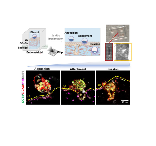
Figure: 3D uterine embryo implantation simulating chip and human embryos (blastocyst or blastoid) implantation process.
(image by Yu Leqian’ Lab)
