Recently, the research team led by Dr. Xingke Yang of the Key Laboratory of Zoological Systematics and Evolution published a paper entitled “Visualization of the 3D structures of small organisms via LED-SIM” in Frontiers in Zoology (2016:13). This research was supported by the National Natural Science Foundation of China (3010300101, 31372239 and 81427802), Research Equipment Development Project of Chinese Academy of Sciences (YZ201509).
Microscopy techniques have been playing an important role in modern zoology. Innovative new techniques (e.g., micro-CT, MRI, CLSM, etc.) have improved our understanding of organismal biology significantly. It is still often challenging to observe internal 3D structures of small animals, despite the use of various Optical and none-Optical techniques.
Dr. Xingke Yang’s team has been collaborated with Dr. Baoli Yao’s lab (State Key Laboratory of Transient Optics and Photonics) in Xi’an Institute of Optics and Precision Mechanics for many years. The LED-SIM method that utilized by the authors is previously developed by Dan et.al. (2013). The LED-SIM is previously applied in imaging of cells and neuron. It has not yet seen application in imaging of small animals which often with hard cuticles and hard to visualize. This research proposed innovative solutions and protocols to obtain high resolution 3-D images in small animals based on ‘SIM’ technique, which can provide a high-speed and high-resolution format. Compared to micro-CT and CLSM, this method of 3-D imaging is with higher resolution in X-Y plane and highly lowered cost of equipment. Although LED-SIM is quite new and rarely used currently, it has a promising future for a large numbers of scientists or labs to access and use due to its high performance and low cost.
Online version of article: http://frontiersinzoology.biomedcentral.com/articles/10.1186/s12983-016-0158-9
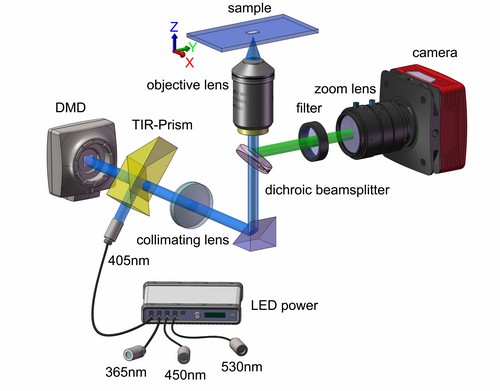
Schematic diagram of the DMD-based LED illumination SIM microscope.
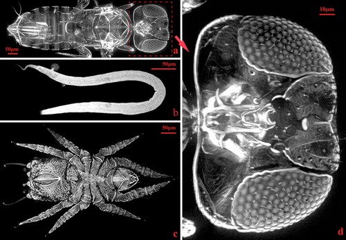
LED-SIM images of different organisms.